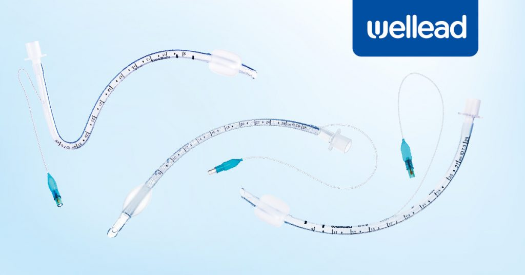
Endotracheal intubation (ETT) is an indispensable resuscitation procedure in emergencies. It involves placing a specially designed tube through the mouth or nose into the trachea (airway/windpipe) to establish an artificial airway.
The endotracheal tube insertion procedure plays an essential role in maintaining patient ventilation, protecting the lower airways, and ensuring adequate gas exchange. It is widely used in intensive care, trauma management, and various surgical procedures. Direct and video laryngoscopy are the two most common methods used for endotracheal intubation.
This article will focus on the key steps and techniques in the ETT insertion procedure.

Why Would a Patient Need Endotracheal Tube Intubation?
Purpose of Endotracheal Tube Intubation
In controlled settings, endotracheal tube intubation secures an airway. It allows tracheal access for ventilatory assistance, anesthetic gas supply, and stomach aspiration prevention. It also lets monitor end-tidal CO2, regulate positive pressure, and prevent dynamic hyperinflation in respiratory compromise. The endotracheal tube insertion procedure includes rapid sequence induction to decrease aspiration risk in trauma or emergent surgery.
When Is the Placement of an Endotracheal Tube Recommended?
- Profound hypoxemia in severe ARDS.
- Neurological injury with compromised airway reflexes.
- Polytrauma with suspected airway obstruction.
- High-grade thermal or caustic airway burns.
- Status epilepticus with ongoing seizure activity.
- Deep sedation for protracted mechanical ventilation.
- Full-thickness facial injury with airway distortion.
- Intraoperative ventilation during neurosurgery.
- Cardiac arrest with ongoing cardiac life support.
- Refractory shock needing aggressive hemodynamic support.
Who Should Not Be Intubated?
Patients with the following conditions shouldn’t be intubated:
- Those with explicit do-not-intubate orders;
- Patients with airway anomalies making endotracheal intubation impossible;
- End-stage disease patients for whom mechanical ventilation offers no survival benefit.
Intubation is risky for patients with high cervical spine instability, maxillofacial trauma, or obstructive lesions beyond conventional laryngoscopy. Forced intubation may worsen their clinical outcomes.
Ethically, respect patient autonomy, weigh adverse events, and recognize that aggressive airway management may conflict with comfort or palliative goals.
Preparation before Insertion
In any endotracheal tube insertion procedure, start with an airway evaluation that assesses mandibular space, Mallampati class, cervical spine mobility, and known anatomical or pathological challenges. Have multiple cuffed and uncuffed endotracheal tubes. Test each cuff with a calibrated syringe for no micro-leaks. Use a laryngoscope system (direct or video). Yet, keep a bougie, fiberoptic scope, or laryngeal mask ready in an unanticipated difficult airway.
Confirm the function of suction, capnography monitors, and oxygen delivery devices. Prepare induction agents (etomidate or propofol) and neuromuscular blockers (succinylcholine or rocuronium) with weight-based dosing and labeled syringes. Have lidocaine or atropine on hand for hemodynamic or airway considerations. To moderate desaturation risks, check IV patency, adjust the patient’s fluid status and blood pressure, and pre-oxygenate with 100% oxygen. Keep vigilance for rapid airway resistance or oxygenation changes throughout the phase.
What Is Endotracheal Tube Insertion Procedure?
How to insert endotracheal tube? During the endotracheal tube insertion procedure, healthcare providers typically perform the following steps:
- Select general anesthesia or local anesthesia based on the patient’s condition.
- Position the patient, optimizing the head position to obtain the best view of the vocal cords, usually in a supine position.
- Open the mouth and insert the laryngoscope into the oral cavity (it can also be inserted into the nasal cavity if necessary). This step involves tools such as a handle, lights, and a dull blade, which can help medical staff guide the tracheal tube.
- Move the tools to the back of the oral cavity to expose the epiglottis and glottis, while avoiding the teeth.
- Slowly insert the endotracheal tube into the patient’s airway along the guide of the laryngoscope.
- Inflate the cuff.
- Remove the laryngoscope.
- Secure the endotracheal tube with tape or other methods to prevent it from shifting.
- Once the endotracheal tube insertion procedure is complete, verify placement. Attach an end-tidal CO2 monitor and observe a waveform for a few consecutive breaths. Auscultate for equal bilateral breath sounds and check for symmetrical chest expansion. It can also be checked by taking an X-ray.
Techniques and Tips for Inserting an Endotracheal Tube
During the endotracheal tube insertion procedure, adjust head elevation so that the external auditory meatus is aligned with the sternal notch. Pre-oxygenate with high-flow oxygen to delay desaturation.
Select a blade (curved or straight) per anatomy and comfort. Then, lift at a 45-degree angle to avoid leverage on teeth.
Confirm placement by end-tidal CO2 waveform and bilateral breath sounds since mainstem intubation or esophageal placement can be tragic. Keeping cuff pressures between 20 and 30 cm H2O can prevent some of these complications.
Seeking a Supply Partner for Endotracheal Tubes?
Well Lead Medical is one of the global suppliers of medical catheters. The production and sales of endotracheal tubes and Foley catheters hold prominent positions in both domestic and international markets.
At Well Lead Medical, we manufacture a wide range of endotracheal tubes with complete specifications, excellent materials, reasonable design, stable quality, and wide application.
Visit our official website to learn more about how our products are designed to reduce lower tracheal trauma during the insertion procedure.

